
Content
Introduction
This video will detail the operative techniques and perioperative considerations employed by the University of Toronto's, Division of General Surgery, for the safe and effective performance of a Laparoscopic Cholecystectomy. This video is directed towards junior surgical residents. It will provide a step-by-step breakdown of the procedure, while highlighting important technical skills and anatomic landmarks.
View Video
Port Sites
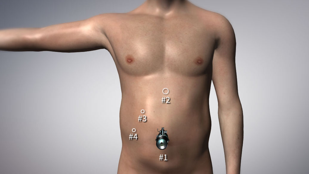
A 10mm trochar placed 2 finger breadths under the xiphoid process, at the midline. A 5mm trocar placed 1-2 finger breadths under the inferior boarder of the right costal margin, at the mid-clavicular line. A 5mm trochar placed inferiorly approximately 1.5 hand breadths away slightly lateral to the level of the previous trochar.
By convention, the umbilical camera port is labelled #1, the 10mm midline trochar #2, the subcostal port #3 and the inferior flank port #4.
Supplementary Material
• WHO Surgical Safety Checklist
Financial Support
This video was generously funded by the University of Toronto, Office of Integrated Medical Education: Educational IT Summer Program.

Recent updates
December 04, 2014
Laparoscopic Cholecystectomy Video launched!
All contents copyright ©2008 - 2013 University Health Network. All rights reserved.
