Lumbar Anatomy Teaching Module
Introduction
The Lumbar Anatomy Teaching Module has been designed to aid students and educators to understand and teach lumbar spinal anatomy by providing them with interactive 3D visuals and complementary text.
Note: This module was developed with Adobe Flash, and Flash is no longer supported by web browsers. However, there is a new Flash emulator called Ruffle which is under development. Currently, the Virtual Spine Lumbar Anatomy, Virtual Bronchoscopy, Cardiac Embryology, Virtual Liver and POPS simulation are the only PIE Flash modules that works with Ruffle, but as development is ongoing, future releases of Ruffle will work with other PIE modules. This will be indicated on the web page for each module.
Ruffle can be installed as an extension to the Chrome browser which will allow these modules to play. This extension can be downloaded from the Chrome Web Store:

Features
- interactive rotateable 3D models of lumbar anatomy
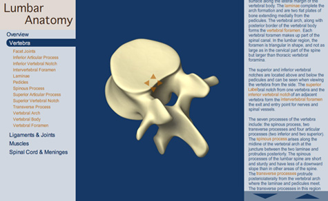
- explores the lumbar anatomy of the vertebra, ligaments, joints, muscles, the spinal cord, and meninges.
References
- Wall EJ, Cohen MS, Abitbol JJ, Garfin SR. Organization of intrathecal nerve roots at the level of the conus medullaris. J Bone Joint Surg Am. 1990; 72(10):1495-1499.
- Wall EJ, Cohen MS, Massie JB, Rydevik B, Garfin SR. Cauda equina anatomy. I: Intrathecal nerve root organization. Spine (Phila Pa 1976). 1990; 15(12):1244-1247.
- Cohen MS, Wall EJ, Brown RA, Rydevik B, Garfin SR. Cauda equina anatomy. II: Extrathecal nerve roots and dorsal root ganglia. Spine (Phila Pa 1976). 1990; 15(12):1248-1251.
- Nowicki BH, Haughton VM. Neural foraminal ligaments of the lumbar spine: Appearance at CT and MR imaging. Radiology. 1992; 183(1):257-264.