Content
 Ultrasound in Trauma: eFAST

Introduction
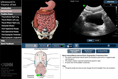
In trauma medicine there is often a need for quick, qualitative assessment of a patient. The Focused Assessment with Sonography in Trauma (FAST) and extended FAST (eFAST) exams are the current standard for rapid evaluation of trauma patients. Training for carrying out eFAST assessment requires practitioners to understand the 3D structures of the body that are seen in the 2D ultrasound image.
This interactive module was created to assist in the teaching and learning of the FAST and eFAST assessments. It shows users how to correctly position their probe and provides examples of both normal and positive ultrasound exams and matches them with 3D visuals to improve understanding of the ultrasound findings.
How to Use the Module
In the normal exam section each eFast view can be selected in the lower left portion of the screen. For each eFast view, the 3D body model, ultrasound probe, and ultrasound plane can be rotated in the horizontal or vertical plane to view it from any angle.
Key features of of the normal section include:
- Rotatable 3D model of relevate anatomy can be rotated in a horizontal and vertical axis. Users can explore the relationship of the probe to the body.
- Remove portion of the 3D body.
Gain a better understanding of the US plane in relationship to the body's structures. - Orient the 3D model probe plane to match the US clip.
Label the structures in the 3D model corresponding to the US clip - Positive exam ultrasound clips.
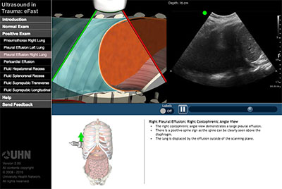
The positive exam section provides only a comparison of the US clip with a cut view of the 3D model with labeling.
Acknowledgements
The development and testing of this module was supported by a grant from the Instructional Technology Innovation Fund (ITIF) at the University of Toronto.
The eFast module was developed by Jean Lin and Jodi Crossingham, with medical content provided by Dr. Massimiliano Meineri and Dr. Alberto Goffi, and with consultation from Dr. Gordon Tait.