Content
Focused Cardiac Ultrasound (FOCUS)
![]() 2020-12-16: This module has been converted from Adobe Flash to HTML5, and is now accessible on tablets and smartphones as well as on desktop computers. At this time, only the core module with the normal views has been converted to HTML5. The views with pathology (pericardial effusion, tamponade, LV and RC dysfunction and hypovolemia) are in the process of being converted and will be available early in the new year (2021).
2020-12-16: This module has been converted from Adobe Flash to HTML5, and is now accessible on tablets and smartphones as well as on desktop computers. At this time, only the core module with the normal views has been converted to HTML5. The views with pathology (pericardial effusion, tamponade, LV and RC dysfunction and hypovolemia) are in the process of being converted and will be available early in the new year (2021).
Click here to open the Focused Cardiac Ultrasound application.
Using an iPad? Click the App Store button to buy the app.
Instructions on updating the TTE FOCUS Views app
Introduction
In the practice of anesthesia, critical care and emergency medicine there is often a need for a quick, qualitative assessment of cardiac function. Focused Cardiac Ultrasound (FOCUS) is the process of carrying out this rapid qualitative assessment by practitioners in these fields. Training for carrying out a FOCUS assessment requires practitioners to understand the 3D structures of the heart that are seen in the 2D TTE image.
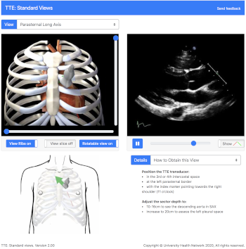
We have created this online interactive module to assist with teaching and learning the assessment of cardiac function with FOCUS. Users can view the TTE recordings for each of the 5 FOCUS views and see a corresponding 3D heart model, ultrasound probe, and ultrasound plane.
- Parasternal long axis
- Parasternal short axis
- Apical four chamber
- Subcostal four chamber
- Subcostal inferior vena cava
How to Use the Module
Each FOCUS view can be selected from a drop-down menu in the upper left of the screen. For each FOCUS view, the 3D heart model, ultrasound probe, ultrasound plane, and rib cage can be rotated in the horizontal or vertical plane to view it from any angle. The rib cage can be removed, the part of the heart above the echo plane can be removed, and the heart model can be oriented so the structures correspond to the TTE image.
Key features of different sections include:
- TTE probe with removable rib cage in the FOCUS views
Add or remove rib cage to locate the TTE probe position in relationship to the heart - Rotatable 3D heart model in all views
Explore the relationship of the imaging plane to the heart by rotating the 3D heart model in the horizontal and vertical planes - Show a section of the 3D heart model corresponding to the TTE clip
Label the structures in the 3D model corresponding to the TTE clip - Introduction to pathology information
Obtain background information to help interpret normal and pathological changes - Animated section of 3D heart model illustrates pathology in TTE clips
Compare normal and pathological animations and TTE clips side by side
Acknowledgements
The development and testing of this module was supported by a grant from the Academic Health Science Centres (AHSC) Academic Funding Plan (AFP) Innovation Fund.
The development of the additional pathology sections of this module was supported by the University of Toronto OIME Summer EIT Program and a grant from the Academic Health Science Centres Academic Funding Plan Innovation Fund. The additional pathology sections was developed by Derek Ng, PhD, MScBMC candidate, under the supervision of Dr. Annette Vegas, Dr. Massimiliano Meineri and Dr. Gordon Tait.
Updating the TTE FOCUS Views app: Users who are updating the TTE FOCUS Views app from the previous version, please delete the app from your iPad and then reinstall the new version from your purchased apps in the App Store. If you simply update the app, there is a problem accessing the normal TTE views. We apologize for the inconvenience, and are working on a solution for this problem.