
Spectral Doppler: Aortic Valve
Obtaining the spectral Doppler
- Identify the aortic valve (AV) in the transgastric views. Either of the two following two-dimensional TEE views can be used:
- Align the continuous wave (CW) cursor through the centre of the AV (figure 1).
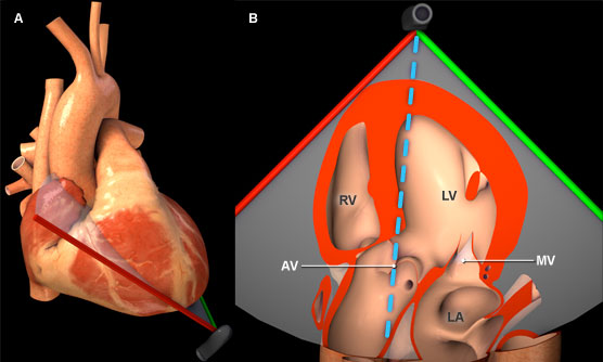
Figure 1: Three-dimensional heart model shown in cross-section to highlight the position of the TEE plane in the deep transgastric view. A) Anterior view of the heart with TEE plane. B) Inferior view of the heart and TEE plane. The blue dotted line represents the orientation of the continuous wave cursor. Key: AV = Aortic valve, LV = Left ventricle, LVOT= Left ventricular outflow tract, MV = Mitral valve, RV = Right ventricle.
Features of aortic valve spectral Doppler
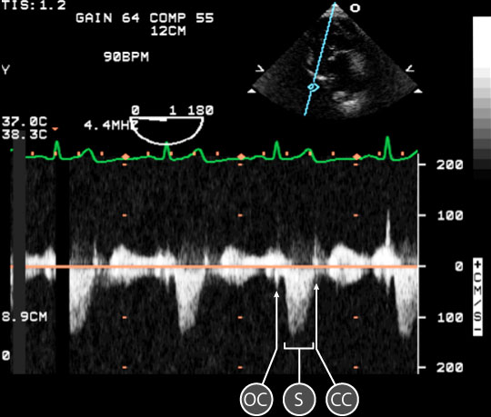
Figure 2: Spectral doppler data acquired for blood flow through the aortic valve. In the upper right, a two dimensional TEE image of the deep transgastric view; the blue line is directed through the tips of the aortic valve leaflets. In the lower half of the image, a spectral doppler trace shows the relationship between red blood cell velocity and time. The baseline is orange. Key: CC = closing click, OC = opening click, S = systolic wave.
- A normal aortic valve spectral doppler trace has a single rounded systolic (S) wave below the baseline, as bloodflow is away from the transducer. The S wave is enclosed within AV opening (OC) and closing clicks (CC) (figure 2).
- The AV wave has a Maximum velocity (VAV) of 80 - 130 cm/sec.
- The AV velocity-time intergral (VTI) is 15 - 25 cm
Physiological variation
- Load dependent
Pathological variation
- AV and subaortic valvular disease