
Spectral Doppler: Mitral Valve
Obtaining the spectral Doppler
- Identify the mitral valve (MV) in the mid-esophageal views. Any of the four following two-dimensional TEE views can be used:
- To assess mitral valve inflow, align the pulsed wave (PW) cursor through the MV and place the sample volume at the MV leaflet tips during diastole (figure 1).
- To assess the MV stroke volume, place the PW sample volume at level of mitral annulus.
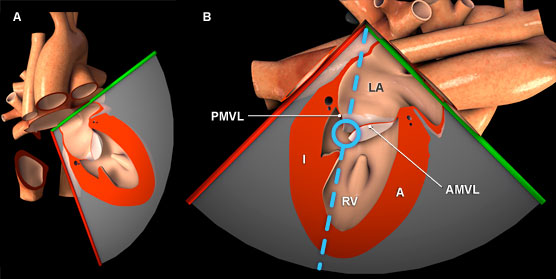
Figure 1: Three-dimensional heart model shown in cross-section to highlight the position of the TEE plane in the mid esophageal two chamber view. A) Anterior view of the heart with TEE plane. B) Anterolateral view of the heart and TEE plane.The blue dotted line represents the orientation of the pulsed wave cursor and the blue circle indicates the position of the sample box at the tip of the mitral valve leaflets. Key: A = Anterior left ventricular wall, AMVL = Anterior mitral valve leaflets, I = Inferior left ventricular wall, LA = Left atrium, LV = Left ventricle, PMVL = Posterior mitral valve leaflet.
Features of mitral valve spectral Doppler
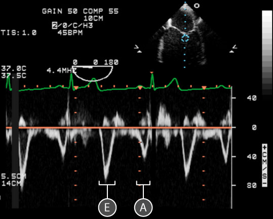
Figure 2: Spectral doppler data acquired for blood flow through the mitral valve. In the upper right, a two dimensional TEE image of the mid esophageal four chamber view; the blue circle indicates the location of the sample volume at the tip of the mitral valve leaflets. In the lower half of the image, a spectral doppler trace shows the relationship between red blood cell velocity and time. The baseline is orange. Key: E = Early diastolic filling wave, A = Atrial kick wave.
- A normal mitral valve inflow spectral doppler trace has two peaks.
- The Early diastolic filling (E) and Atrial kick (A) waves are seen below the baseline as the direction of blood flow is away from the transducer (figure 2).
- The E wave has Maximum velocity (Emax) of 60 - 80 cm/sec
and a Deceleration time (Edecel) of 160 - 240 msec - The A wave has a Maximum velocity (Amax) of 20 - 40 cm/sec
and A duration (TMADur) of 150 – 240 msec. - The normal E/A Ratio is 1 – 1.5
Physiological variation
- Aging (↓ E, ↑ A)
- Pre/afterload
- Tachycardia (E and A wave fusion and ↓ E/A ratio)
- Bradycardia causes ↑ E/A ratio
- Respiratory variation (↑ E, ↑ A during spontaneous expiration)
- Valsalva (↓ preload with ↓ E, ↓ A and ↑ E/A)
Pathological variation
- Diastolic dysfunction
- Arrhythmias
- Mitral and aortic valve disease
- Pericardial disease
- Cardiomyopathies