
Spectral Doppler: Hepatic Veins
Obtaining the spectral Doppler
- Identify the hepatic vein (HV) in the following two-dimensional TEE view:
- To aid in the identification of the HV, apply colour Doppler using a low velocity scale (< 40 cm/sec).
- To assess hepatic vein blood flow velocity align the pulsed wave (PW) cursor through the HV and place the sample volume in the centre of the HV (figure 1).
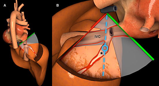
Figure 1: Three-dimensional heart model shown in cross-section to highlight the position of the TEE plane in the modified transgastric hepatic vein view. A) Right posterior view of the heart and liver with TEE plane. B) Posterior view of the heart and liver. The blue dotted line represents the orientation of the pulsed wave cursor, and the blue circle indicates the position of the sample box in the hepatic vein. Key: * = Hepatic vein, IVC = Inferior vena cava.
Features of hepatic vein spectral Doppler
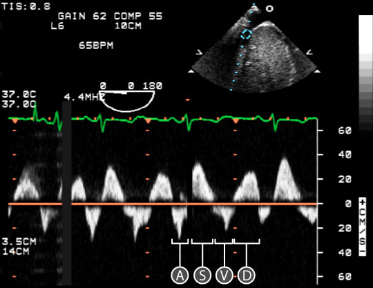
Figure 2: Spectral doppler data acquired for blood flow through the hepatic vein. In the upper right, a two dimensional TEE image of the modified-transgastric hepatic vein view; the blue circle indicates the location of the sample volume within the hepatic vein. In the lower half of the image, a spectral doppler trace shows the relationship between red blood cell velocity and time. The baseline is orange. Key: A = Atrial systolic flow wave, D = Ventricular diastolic flow wave, S = Ventricular systolic flow wave, V = End ventricular systolic flow wave.
- A normal hepatic vein spectral Doppler trace has four peaks (figure 2):
- A wave - a result of retrograde atrial systolic flow. It is seen below the baseline as blood flow is away from the transducer.
- S wave - a result of antegrade ventricular systolic flow. It is seen above the baseline as blood flow is towards the transducer.
- V wave - a result of retrograde end-ventricular systolic flow. It is seen below the baseline as blood flow is away from the transducer.
- D wave - a result of antegrade ventricular diastolic flow. It is seen above the baseline as blood flow is towards the transducer.
Physiological variation
- Respiratory variation (↑ S, ↑ D with spontaneous inspiration)
Pathological variation
- Tricuspid valve disease
- Right sided heart disease
- Cardiomyopathy
- Pericardial disease