
Cardiac Valves
3D animated models
for teaching function and pathology of cardiac valves
We are in the process of developing 3D animated models of a heart and cardiac valves which can be viewed online as visual aids for teaching normal and abnormal cardiac function. These models will allow the user to view the valve from any perspective and animate the opening and closing of the valve. The models depicting normal structure and function will be modified to illustrate various pathological conditions.
Preliminary models
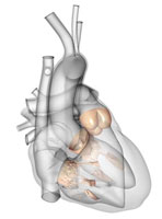
Please click here or on the heart above
to view
a larger rotatable version of the same model.
Below are links to examples of preliminary models of the aortic, pulmonary, mitral and tricuspid valve. Click on an image to open an interactive version of the valve in the image. The valves in the images are preliminary models intended to demonstrate the technology. Final models will be further developed and refined after extensive consultation with cardiac surgeons, cardiologists and anesthesiologists.


We will also animate the flow of blood through the valve using “smoke streams” to visualize the path taken by the blood to show laminar flow as well as the turbulence and vortices created by obstructions.
These models will be made freely available on a Web site, providing educators around the world with a unique teaching resource. It will also provide a self-study resource for residents and Fellows in cardiac surgery, cardiology and anesthesiology.