
Virtual TEE website
Introduction
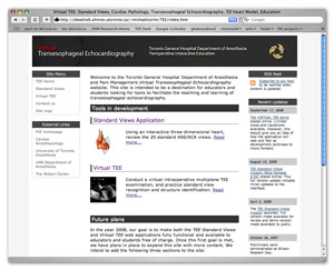
Click on the image to open the Virtual TEE website in a new window.
Visual Interactive Resource for Teaching, Understanding And Learning Transesophageal Echocardiography (VIRTUAL TEE) is an online teaching aid for TEE. It is be made up of a variety of teaching modules meant to supplement traditional teaching methods and tools.
The TEE modules developed to date are: Standard Views, Alternative Views, Colour Doppler, Spectral Doppler, Virtual TEE, TEE Simulation and Pathology Synopses.
Standard Views 3D module
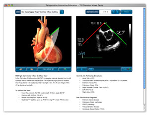
Click on the image to open the TEE standard views page on the Virtual TEE website.

The primary challenge in learning TEE is translating the two dimensional echocardiographic image into a visualization of the three-dimensional (3D) structure of the heart. The TEE Standard Views module provides a learning environment where users can view all of the 20 standard TEE positions using two visualization methods simultaneously: (1) a rotatable 3D heart model that includes an echocardiographic plane and (2) the associated TEE clip. The 3-D heart model and echo plane can be rotated, helping students to relate the echocardiographic image to the structures of the heart. Students are also able to remove the part of the heart above the echo plane, revealing the internal structures of the heart that correspond to the TEE image. This resource can be used both by educators for teaching small group sessions and by students for self-study.
This module is available in the following languages:
- English
- Chinese (中文)
- French (Français)
- Italian (Italiano)
- Japanese (日本語)
- Russian (русский язык)
- Polish (polszczyzna)
- Spanish (Español)
The development and testing of this project is possible thanks to the generous support of the University of Toronto Instructional Technology Courseware Development Fund.
Open the Standard Views 3D Module page on the VIRTUAL TEE website in a new window
Standard Views Guide
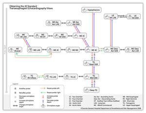
Click on the image to open the Standard Views: Guide Sheet page on the VIRTUAL TEE website.
The Standard Views Guide Sheet is a resource designed to help clinicians learn the relationships between the Standard TEE views. The guide comes in two flavours: online interactive and printable electronic document.
Open the Standard Views Guide page on the VIRTUAL TEE website in a new window.
VIRTUAL TEE module
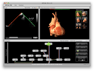
Click on the image to open the VIRTUAL TEE page on the VIRTUAL TEE website.
VIRTUAL TEE is an online teaching aid designed for use by educators and students of two-dimensional TEE. It is intended to help novice and experienced echocardiographers develop a better understanding of the spatial relationship between ultrasound plane and heart, as well as provide new echocariographers with a structured and logical description of the relationships between the American Society of Echocardiographers (ASE) standard views. Students are able to conduct a virtual TEE study, advancing and rotating the probe and image plane and viewing the resulting TEE images as they conduct the study. They are challenged to position the probe to achieve each of the 20 standard views.
Open VIRTUAL TEE module on the VIRTUAL TEE website in a new window.
Alternative Views
Click on the image to open the Alternative Views module on the VIRTUAL TEE website.
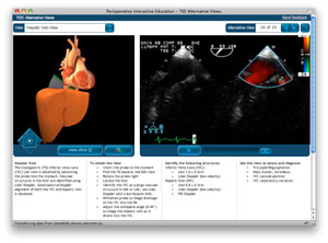
The TEE Alternative Views module provides a learning environment where users can explore 19 non-standard but often used 2D TEE views using two visualization methods simultaneously: (1) a rotatable 3D heart model that includes an echocardiographic plane and (2) the associated TEE clip.
Open the Alternative Views module page on the VIRTUAL TEE website in a new window.
TEE Simulation: Introdction to Probe Manipulation
Click on the image to open the TEE Simulation module on the VIRTUAL TEE website.
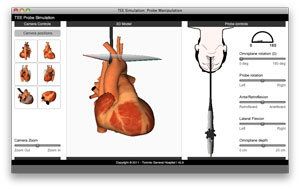
The TEE Simulation module simulates the actions you can take to adjust the position of the TEE probe and the ultrasound plane, and shows you the resulting position of the ultrasound plane in relation to a 3D model of the heart.
Open the TEE Simulation module page on the VIRTUAL TEE website in a new window.
Spectral Doppler
Click on the image to open the Spectral Doppler module on the VIRTUAL TEE website.
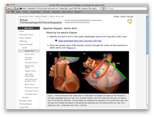
This module demonstrates the assessment of structures using spectral Doppler during the standard 2D examination. The normal spectral waveform for each valvular and vascular structure is accompanied by an anatomical slice of a 3D heart model displaying the spectral beam alignment. Video clips indicate the useful 2D views for optimal Doppler alignment.
Open the Spectral Doppler module page on the VIRTUAL TEE website in a new window.
Colour Doppler
Click on the image to open the Colour Doppler module on the VIRTUAL TEE website.
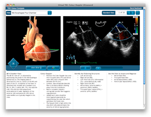
The application of colour Doppler imaging is routinely performed over cardiac valves, vascular and other pathological structures to assess blood flow direction and velocity. This module demonstrates which structures should be viewed using colour Doppler during the standard 2D examination.
Open the Colour Doppler module page on the VIRTUAL TEE website in a new window.
Pathology
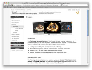
Click on the image to open the Pathology module on the VIRTUAL TEE website.
The Pathology Synopsis Section has three synopses: calcific aortic stenosis, sinus of Valsalva aneurysm (SOVA) and cardiac myxoma. Our pathology synopsis section is available for browsing on the iPhone. Click here to open the iPhone version of the SOVA synopsis.
Open the Pathology module page on the VIRTUAL TEE website in a new window.