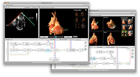
VIRTUAL TEE
Visual Interactive Resource for Teaching, Understanding And Learning Transesophageal Echocardiography (VIRTUAL TEE) is an online teaching aid designed for use by educators and students of two-dimensional TEE. It is intended to help novice and experienced echocardiographers develop a better understanding of the spatial relationship between ultrasound plane and heart, as well as provide new echocariographers with a structured and logical description of the relationships between the American Society of Echocardiographers (ASE) standard views.
Features
- Video clips of twenty standard TEE views, as well as clips of the transitional movements between them.
- A three-dimensional model of the heart demonstrating ultrasound probe and plane position.
- The ultrasound plane is animated so that its motion echoes the movement in the video clips.
- The heart can be viewed from one of four views (anterior, superior, left, right) at all times, and can be rotated around two axes when the plane is stationary at any of the standard views.
- At each standard view, a wedge of heart tissue can be hidden to reveal the internal structures that the echo plane passes through.
- A graphic that maps the relationship between the standard views and also serves as a tool for navigating to desired views. A blue, pie-slice-shaped TEE icon hovers over the map providing the user with feedback about their current position in the virtual examination.
Contributors
- Dr. Massimiliano Meineri
Staff anesthesiologist
Toronto General Hospital
Department of Anesthesia and Pain Management
- Dr. Annette Vegas
Staff anesthesiologist
Toronto General Hospital
Department of Anesthesia and Pain Management
- Michael Corrin
Programming & Design
Toronto General Hospital
Department of Anesthesia and Pain Management

